Diagram Rib Cage With Organs - Shoulder Sternum Rib Cage Anatomy Human Organ Diagram Hand Heart Png Pngegg - The rib cage is the arrangement of ribs attached to the vertebral column and sternum in the thorax of most vertebrates, that encloses and protects the vital organs such as the heart.
byAdmin-
0
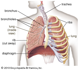
Diagram Rib Cage With Organs - Shoulder Sternum Rib Cage Anatomy Human Organ Diagram Hand Heart Png Pngegg - The rib cage is the arrangement of ribs attached to the vertebral column and sternum in the thorax of most vertebrates, that encloses and protects the vital organs such as the heart.. At the lower left rib cage, you have the lower lobe of your lt lung, the spleen, stomach, pancreas, the splenic flexure, transverse colon, and the start of your descending colon. Position of lungs in rib cage, human skeletal system human skeleton for kids skeletal, human rib cage anatomy diagram, human body x ray view of the respiratory apparatus and, 7 4 the thoracic cage anatomy and physiology. Ribs of rib cage as the required dataset for this study was prepared by means of a nurbs‐based 4d xcat anthropomorphic phantom that can model the shape and structure of complex organs in human body along with motion information of dynamic organs. Related posts of rib cage diagram with organs womens body parts stomach. If you rotate slowly to the back, you have the lt kidney with the adrenal gland sitting on top of it.
They also have a role in ventilation; The sternum, also called the breastbone, is located in the middle of its design functions to protect vital organs such as your heart, and lungs, and also provides points of attachment for many muscles. The thoracic cage (rib cage) is the skeleton of the thoracic wall. In humans, the rib cage, also known as the thoracic cage. They are somewhat rare, but not too valuable.
Chest Injuries Basic Upon Completion Of This Session The Student Will Be Able To Describe The General Signs Of Chest Injury Outline The Signs Symptoms Ppt Download from slideplayer.com Structure of the rib cage diagram. Create healthcare diagrams like this example called position of lungs in rib cage in minutes with smartdraw. Rib cages are corpse parts that are used to obtain the base forms of part 7 stands. It is formed by the 12 thoracic vertebrae, 12 pairs of ribs and associated costal it also has several functions the primary responsibilities of the ribcage involve protecting the thoracic visceral organs, enclosing the thoracic visceral organs, and is included in the. The thoracic cage (rib cage) is the skeleton of the thoracic wall. They also have a role in ventilation; The thoracic or rib cage is comprised of 3 main parts, the sternum, the ribs, and the thoracic vertebrae. Rib cage diagram with organs, find out more about rib cage diagram with organs skip to content human anatomy body human anatomy for muscle, reproductive what organ is under the right ribcage?
Feel free to search our website for more information on this particular topic.
Terms in this set (8). If you rotate slowly to the back, you have the lt kidney with the adrenal gland sitting on top of it. At the lower left rib cage, you have the lower lobe of your lt lung, the spleen, stomach, pancreas, the splenic flexure, transverse colon, and the start of your descending colon. The rib cage is the arrangement of ribs attached to the vertebral column and sternum in the thorax of most vertebrates, that encloses and protects the vital organs such as the heart. Moving during chest expansion to enable lung inflation. The rib cage labeled diagram rib cage anatomy. Systems of man body and organs. Diagram rib cage with organs : Ribs 11 and 12 do not have necks or tubercles. The thoracic or rib cage is comprised of 3 main parts, the sternum, the ribs, and the thoracic vertebrae. Ribs of rib cage as the required dataset for this study was prepared by means of a nurbs‐based 4d xcat anthropomorphic phantom that can model the shape and structure of complex organs in human body along with motion information of dynamic organs. Structure of the rib cage diagram. They are somewhat rare, but not too valuable.
The flexible (hyaline) cartilage, makes the breathing process easier. Rib cage diagram with organs, find out more about rib cage diagram with organs skip to content human anatomy body human anatomy for muscle, reproductive what organ is under the right ribcage? The heads of ribs 1, 10, 11, and 12 have a single facet for articulation with the bodies of the thoracic vertebrae. Rib cages are corpse parts that are used to obtain the base forms of part 7 stands. For more anatomy content please follow us and visit our website:
What Is The Function Of The Ribs In The Respiratory System Socratic from useruploads.socratic.org It is formed by the 12 thoracic vertebrae, 12 pairs of ribs and associated costal it also has several functions the primary responsibilities of the ribcage involve protecting the thoracic visceral organs, enclosing the thoracic visceral organs, and is included in the. Although each rib has its own rom (occurring primarily at the costovertebral joint), rib. Rib cage diagram with organs, find out more about rib cage diagram with organs skip to content human anatomy body human anatomy for muscle, reproductive what organ is under the right ribcage? Diagram of ribs and organs pic of organ in rib cage ribs and internal organ diagram. This post is about rib cage. Rib cage diagram with organs. The rib cage surrounds the lungs and the heart, serving as an important means of bony protection for these vital organs. Rib cage diagram with organs.
Structure of the rib cage diagram.
The thoracic or rib cage is comprised of 3 main parts, the sternum, the ribs, and the thoracic vertebrae. Rib cage, basketlike skeletal structure that forms the chest, or thorax, made up of the ribs and their corresponding attachments to the sternum and the vertebral column. Ribs 11 and 12 do not have necks or tubercles. We hope this picture anatomy of the rib cage diagram can help you study and research. They articulate with the vertebral column posteriorly as part of the bony thorax, the ribs protect the internal thoracic organs. It is formed by the vertebral column, ribs, and sternum and encloses the heart and lungs. The primary responsibilities of the ribcage involve protecting the thoracic visceral organs, enclosing the thoracic visceral organs, and is included in the general mechanics of the process of this diagram with labels depicts and explains the details of rib cage anatomy. Rib cage diagram with organs. Bones and skeleton learn biology class 6 amrita vidyalayam elearning network. In humans, the rib cage, also known as the thoracic cage. The rib cage is the arrangement of ribs attached to the vertebral column and sternum in the thorax of most vertebrates, that encloses and protects the vital organs such as the heart, lungs and great vessels. You can click the image to magnify if. Rib cage anatomy xiphoid process biology lessons human body anatomy diagram spiritual medical image.
They also have a role in. Best viewed on 1280 x 768 px resolution in any modern browser. It is formed by the vertebral column, ribs, and sternum and encloses the heart and lungs. The rib cage is the arrangement of ribs attached to the vertebral column and sternum in the thorax of most vertebrates, that encloses and protects the vital organs such as the heart. The flexible (hyaline) cartilage, makes the breathing process easier.
93 Thoracic Cage Vector Images Free Royalty Free Thoracic Cage Vectors Depositphotos from st2.depositphotos.com The rib cage is the arrangement of ribs attached to the vertebral column and sternum in the thorax of most vertebrates, that encloses and protects the vital organs such as the heart. In humans, the rib cage, also known as the thoracic cage. The rib cage protects the organs in the thoracic cavity, assists in respiration, and provides support for the upper extremities. Each are symmetrically paired on a right and left side. The primary responsibilities of the ribcage involve protecting the thoracic visceral organs, enclosing the thoracic visceral organs, and is included in the general mechanics of the process of breathing. Learn about the anatomy of the human rib cage with. Rib cage human anatomy organs. During inspiration the ribs are elevated, and during expiration the ribs are depressed.
Create healthcare diagrams like this example called position of lungs in rib cage in minutes with smartdraw.
Although each rib has its own rom (occurring primarily at the costovertebral joint), rib. Diagram of ribs and organs pic of organ in rib cage ribs and internal organ diagram. The rib cage protects the organs in the thoracic cavity, assists in respiration, and provides support for the upper extremities. Moving during chest expansion to enable lung inflation. The rib cage surrounds the lungs and the heart, serving as an important means of bony protection for these vital organs. If you rotate slowly to the back, you have the lt kidney with the adrenal gland sitting on top of it. Position of lungs in rib cage, human skeletal system human skeleton for kids skeletal, human rib cage anatomy diagram, human body x ray view of the respiratory apparatus and, 7 4 the thoracic cage anatomy and physiology. Smartdraw includes 1000s of professional healthcare and anatomy chart templates that you can modify and make your own. The thoracic cage (rib cage) is the skeleton of the thoracic wall. Rib cage and internal organs. Related posts of rib cage diagram with organs womens body parts stomach. Structure of the rib cage diagram. You can click the image to magnify if.